Molecular Horizons Second Annual Light Microscopy Image Competition
MicroWorld in Motion Winners (3D image and video)
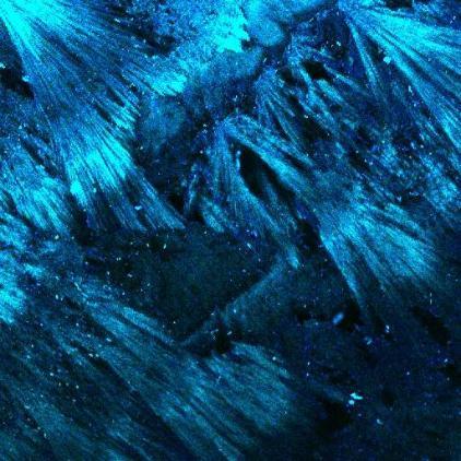
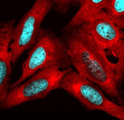
- 1st Place | Luke McAlary: Liquid Phase Separation of DDX4 protein
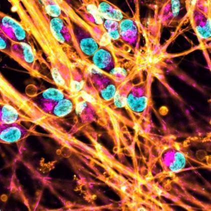
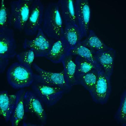
- 2nd Place | Benjamin Genenger: Live cell imaging of co-culture skin cancer spheroids
Description
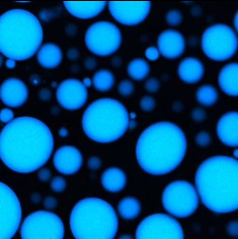
Liquid Phase Separation of DDX4 protein
Supervisor
Watch the Liquid Phase Separation of DDX4 protein on YouTube
Description
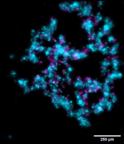
Live cell imaging of co-culture skin cancer spheroids. The patient-derived skin cancer cell line, UW-CSCC2 (magenta), and dermal fibroblasts (cyan) were seeded in ultra-low attachment plates to facilitate spheroid formation. Z-stacks of live spheroids were taken every 2h45min for 60h. This revealed the formation of a cancer shell around a fibroblastic core. Investigation of other combinations of fibroblasts and cancer cell lines indicate the formation of distinctive spatial architectures for each combination (not shown).
Supervisor
Watch live cell imaging of co-culture skin cancer spheroids on YouTube