April 22, 2020
SCIT medical imaging analysis recently published in the top-tier outlet
According to the statistics of the Australian Institute of Health and Welfare, by average, about 1750 people are diagnosed with brain cancer each year in Australia, which means roughly one brain cancer diagnosed every five hours. Also, in Australia brain cancer is the most life-threatening cancer for children and people under 40.
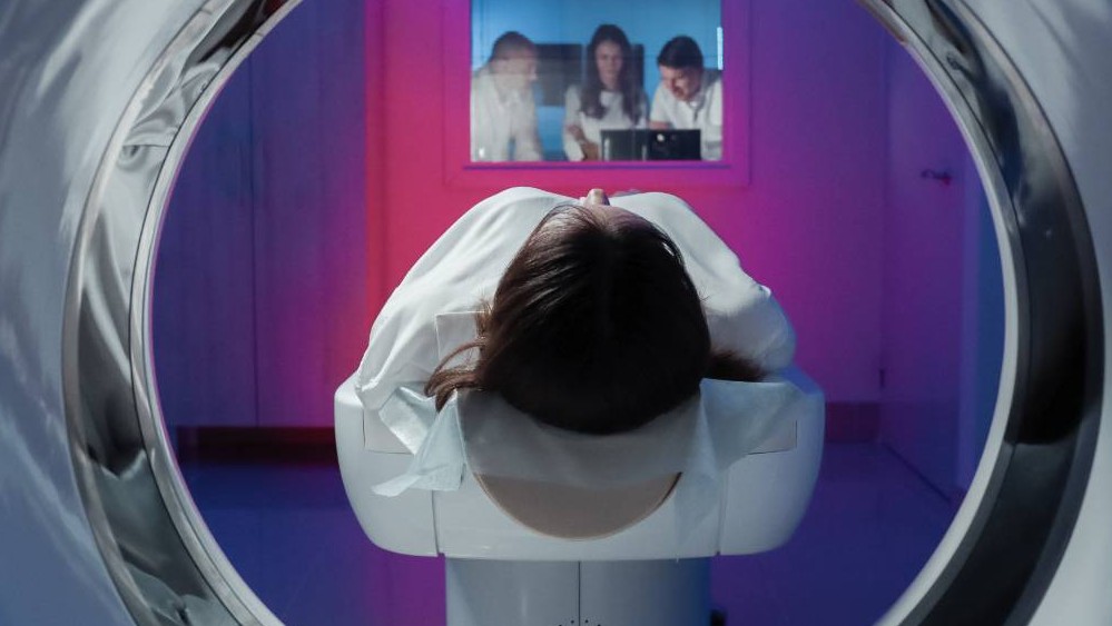
For effective diagnosis and treatment of brain cancers, obtaining the location and appearance information on brain tumours plays a key role. Such information is usually acquired by the non-invasive magnetic resonance imaging (MRI). MRI produces multiple modalities of images and each of them shows unique soft tissue contrast. Collectively using these multi-modality MR images can achieve better diagnosis and treatment of brain cancers.
Nevertheless, due to modality missing and modality inconsistency across clinical centres, the high demand of collectively using multiple MR imaging modalities for analysis is not always met in practice, which could affect the quality of diagnosis and treatment.
A team of researchers from the University of Wollongong, University of Sydney, CSIRO, and Nanjing University have recently developed advanced algorithms to synthesize new modality images based on available modality images. By this cross-modality MR image synthesizing technique, the issues of modality missing and inconsistency can be well mitigated, enabling the collective use of multi-modality MR images as desired. An example of their work is to synthesize fluid-attenuated inversion recovery (FLAIR) MR images from T1-weighted MR images, so that brain tumours could be better viewed for diagnosis and treatment.
Compared with existing cross-modality MR image synthesizing techniques, the methods proposed by this team better overcome the issues of slice discontinuity and blurring observed in synthesized images. To achieve this, one of their work utilizes the edges that contain critical textural information from available modality images and integrate the edge maps into the synthesis model to boost synthesis quality; another work caters for the individual characteristic of each image and preserves this information by learning from the neighbours of each given image.
The proposed methods have been validated on two benchmark data sets in MRI analysis research which contain brain lesions. By comparing them with a set of state-of-the-art image synthesis methods, the effectiveness of the proposed methods is verified by consistently higher values with respect to both qualitative and quantitative measures.
Their work has been published in IEEE Transactions on Medical Imaging, which is a top-tier journal on medical imaging analysis. With one of their papers, the UOW PhD student researcher on this project, Ms Biting Yu, under the supervision of Associate Professor Lei Wang in School of Computing and Information Technology, participated and won the Dolby Scientific Paper Competition in 2019.