August 30, 2017
Teeth reveal last months of mother and child who lived 27,000 years ago
Synchrotron light analysis of dental enamel gives insight into early human life
A team of researchers has used synchrotron light to study the teeth of a prehistoric human foetus and learn about the life and death of the child and its mother, who lived 27,000 years ago.
The team was led by Alessia Nava from Sapienza University of Rome and the Museum of Civilisations of Rome, and included Professor Claudio Tuniz from the University of Wollongong (UOW), as well as researchers from Elettra-Sincrotrone in Trieste, the “Abdus Salam” International Centre for Theoretical Physics in Trieste (ICTP), the Fermi Centre in Rome, and the University of Bari. The study has been published in the journal Scientific Reports – Nature.
Dental enamel is a biological archive that constantly tracks periods of good and bad health while forming. Prenatal enamel, which grows during intrauterine life, reveals the mother's history as well.
“Teeth represent a sort of black box, able to record much information: what type of hominids they belong to (the Neanderthal’s enamel, for example, is generally thinner than the one of a Homo sapiens), the kind of diet they followed, their age, their biological evolution,” said Professor Tuniz, a Visiting Professorial Fellow with UOW’s Centre for Archaeological Research, who is also affiliated with the Fermi Centre and ICTP.
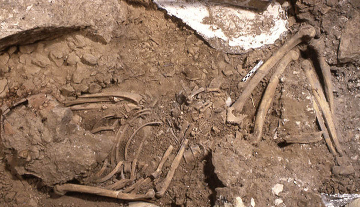
The skeleton of the young woman and her foetus were found in 1991 in the Ostuni 1 burial site in southern Italy. The woman was 20 years of age or younger and in the advanced stages of pregnancy at the time of her death.
The researchers analysed three still forming incisors belonging to the foetus using synchrotron x-ray microtomography, which made it possible to carry out a virtual histological analysis of the fossil teeth, revealing the finest structures of the dental enamel in a non-destructive way.
This is the first time researchers have been able to reconstruct the life and death of an ancient foetus and, at the same time, shed light on its mother's health.
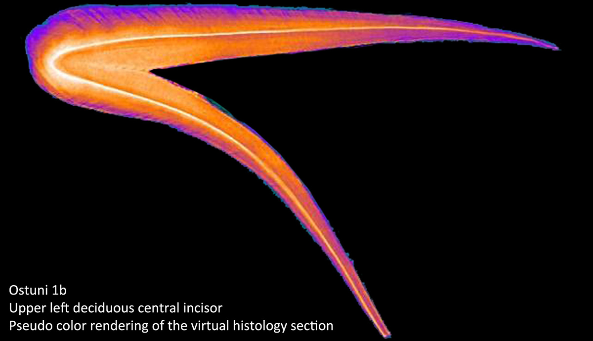
The virtual histological analysis showed that the mother’s and baby's death occurred between the 31st and 33rd gestational weeks. Moreover, three severe physiological stresses affected both individuals during the last two and a half months of life, as highlighted by stress markers visible at microscopic level in the dental enamel.
“Synchrotron light can illuminate the human deep past. In this case advanced physics tools and methods helped to reconstruct details on the last months of life of a pregnant woman from southern Italy 27,000 years ago,” Professor Tuniz said.
“This is the oldest evidence of the price that women had to pay during human evolution. The larger brains that gave humans their cognitive capacities implied a much more risky delivery process.”
"Virtual histological assessment of the prenatal life history and age at death of the Upper Paleolithic fetus from Ostuni (Italy)" by Alessia Nava, Alfredo Coppa, Donato Coppola, Lucia Mancini, Diego Dreossi, Franco Zanini, Federico Bernardini, Claudio Tuniz and Luca Bondioli is published online on Scientific Reports - Nature.
Main image: The Ostuni 1 burial in the Santa Maria di Agnano cave during excavation. Photograph by E.Vacca