August 12, 2015
Low-cost MRI system puts health care in reach for vulnerable communities
A low-cost and easy-to-operate MRI system could help save millions of lives in developing countries.
Magnetic resonance imaging (MRI) uses magnetic fields and radio waves to provide clinicians with three-dimensional scans of body organs and soft tissue sections, providing vital information in diagnosing disease and injury to the brain, spine, muscles and joints.
Yet, the sophisticated machines cost in the vicinity of $1-3 million, as well as the ongoing operation and maintenance costs. Even in developed countries, scans cost several thousands dollars and those partially covered by government rebates can leave patients hundreds of dollars out of pocket.
Researchers from the University of Wollongong have developed a system that could reduce medical costs for people living in regional areas.
Posted by WIN News Illawarra on Tuesday, 18 August 2015
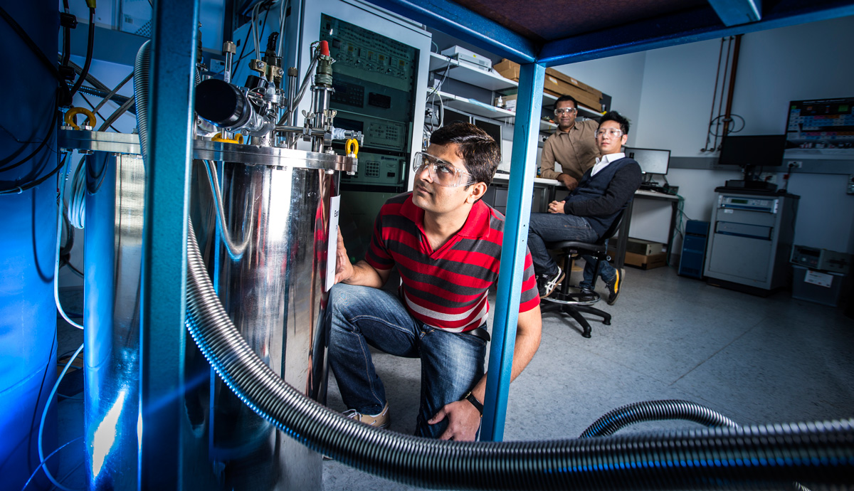
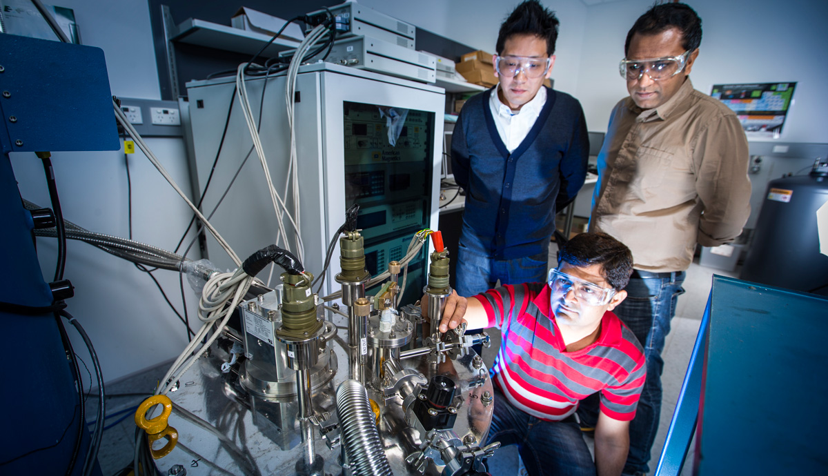
“Diagnostic imaging is critical for quality health care, yet most current imaging devices used for diagnosis and treatment planning are expensive to operate and maintain, putting this treatment of out of reach of millions of people around the world,” Institute for Superconducting and Electronic Materials (ISEM) PhD student Dipak Patel said.
“Patients regularly face long waiting times for scans due to a lack of MRI systems and in the developing world there are many countries that do not have a single MRI unit or have only very old systems in large cities. For a person with a serious injury or illness, it’s not possible to travel hundreds of kilometres to get a diagnosis, let alone be able to afford it.”
A major contributor to the cost of MRI systems is constant cooling of a key part of the system - the magnet - to remain within a set temperature range. To achieve this, the magnet is immersed in a helium ‘bath’. Helium is very expensive and there are concerns about global supply shortages,” Dipak said.
“If a helium-cooled MRI unit has a power outage, the helium boils off, or evaporates, as the magnet heats up. It’s very expensive to refill the cooling chamber and this leads to lengthy downtime during which the system cannot be operated.”
To get around the helium issue, researchers at ISEM, led by ARC Future Fellow Associate Professor Jung Ho Kim, have developed a prototype for a new magnet and cooling system that will enable them to build a low-cost and easy-to-operate MRI system.
The solution uses a new superconducting magnet based on cheap and readily available chemical elements magnesium and boron. The compound wire can be wound into coils, significantly reducing weight compared to conventional magnets and making possible mobile or benchtop MRI systems that could be driven to remote areas to provide essential diagnostics services.
A second critical factor is the magnet operates at a higher temperature than conventional magnets. This makes possible the use of cheap solid nitrogen to maintain the magnet’s critical working temperature, which means it can stay cool even during power interruptions.
“The great thing about solid nitrogen is its thermal stability. It’s a little bit like packing an Esky with ice and leaving it closed. Your drinks can stay cold for a long time. In the case of a power outage, the solid nitrogen stays cool and clinicians can continue to work until power is restored.”
A second challenge the researchers overcame, one that has held back this type of superconducting magnet since its emergence as a candidate material in 2001, was finding a method of joining the superconductor sections.
“For good quality images and lower operating costs the magnet is energized with a charge or electric current then the two ends of the magnet are joined to create a closed loop,” ARC DECRA Senior Fellow and project co-supervisor Dr Shahriar Hossain explained.
“Without a joint the MRI unit must have constant direct current power to maintain the magnetic field, which involves additional costs in equipment and electricity, further driving up the cost of scans for patients.”
The compound decomposes at high temperature, ruling out fusing the sections together. The method Dipak and the team have developed involves cold-joining the wire ends then heat treating the coil in a furnace so that is retains its superconducting properties.
The joining technique was recently published in the journal Superconductor Science and Technology.
Associate Professor Kim said the team have validated the prototype in the lab and are now working on a method of demonstrating it can be mass-produced to meet the demands of MRI manufacturers.
“We’ve shown that our joining method meets the technical needs for use in MRI. If we can get a industry partner we could take this to the next level in providing healthcare services for millions of people worldwide,” he said. “After 30 years of commercial production, superconducting MRI systems have reached a state of maturity. The demands of the healthcare industry are for high efficiency, low cost and reliable systems.”