Molecular Cell Biology

Molecular cell biology

By applying a diverse array of molecular and cellular approaches, we study the operating principles of the process that support life. By integrating biochemistry, molecular biology and cellular biology with advanced imaging and analysis technology our researchers seek a mechanistic understanding of the molecular and cellular pathways related to disease.

Research groups

Our research integrates technologies in human pluripotent stem cell (hPSC) biology, neuroscience, and bioengineering to create cellular models of the human nervous system. Specifically, we work on establishing new techniques for differentiating hPSCs into various neural lineages, such as dorsal root ganglia sensory neurons, cortical neurons and auditory neurons. Our work plays a pivotal role in advancing our understanding of human neurodevelopment and has impact on regenerative medicine applications for neurological disorders, specifically Friedreich's ataxia (FA) and hearing loss.

Our laboratory has also developed valuable tools for collaborative research in modelling neurodegenerative conditions. This includes generation of NGN2 transgenic hPSC lines, which allows for rapid conversion of neurons in vitro (following the protocol of Fernandopulle et al.). Our transgenic hPSC lines includes FA induced pluripotent stem cell lines (iPSC) and their corresponding isogenic control iPSC lines, that were developed by Dr Marek Napierala (owned by University of Alabama, USA). The transgenic cell lines have been applied to study disease mechanisms underlying FA and to test potential therapeutic interventions, including candidate pharmaceutical compounds and gene therapy.

View Scholars page

The Dowton group is directed at understanding how DNA molecules evolve with particular interest in the mitoochondrial genome, which has remarkably little non-coding DNA.  For example, protein-coding regions account for 70% of the mitochondrial genome in humans, but only 1% of the nuclear genome.  Genes are sandwiched together, often with only a few non-coding nucleotides between them.  This has resulted in remarkable stability in the arrangement of genes, as any gene movement is likely to disrupt the function of a neighbouring gene; many genes are in precisely the same position that they were in hundreds of millions of years ago.  Our research has focussed on one lineage of animals that have broken this trend.  The Hymenoptera (ants, bees and wasps) have mitochondrial genomes whose genes change positions relatively frequently.  By sequencing related hymenopteran mitochondrial genomes, we can identify the sorts of changes that have occurred, and better understand the fundamental mechanism of mitochondrial gene rearrangement.

View Professor Mark Dowton's Scholars page

Contact mdowton@uow.edu.au for more information.

A key focus of the lab is to better understand the biological processes that contribute to the invasion and spread of cancer around the body, a process known as metastasis, which is the leading cause of cancer-related deaths. We have made significant progress towards identifying the role of a tissue-degrading system integral to cancer metastasis with multiple publications. We know that high levels of a key component of this system, the enzyme uPA, results in patients with many types of cancer having a greater chance of developing metastasis. As a cell surface protein, uPA represents an accessible druggable target for stopping the metastatic process. To this end, we are utilising a naturally occurring inhibitor of uPA called PAI-2 (Figure 1). Another project is repurposing the old drug amiloride into a potent dual-targeting inhibitor of two well-established cancer biomarkers including uPA.

Our lab also utilises patient tumours for mutational and expression analyses of biomarkers of metastasis and actionable therapeutic targets in skin cancer (Figure 2). Patient-derived cultures and xenografts have been established from fresh tumours and isolated CTCs, as models for targeted genetic manipulation and drug responsiveness studies.

The Ranson lab also contributes to on-going collaborations investigating the role of the uPA system in protein misfolding diseases such as pregnancy preeclampsia and in streptococcal pathogenesis.

View Senior Professor Marie Ranson’s Scholars page

Contact mranson@uow.edu.au for more information.

The Immunology and Cell Signalling Group focuses on extracellular signalling pathways between immune cells in the context of health and disease in humans and companion animals. A key focus of the group is the study of signalling pathways mediated by extracellular ATP and purinergic receptors (mainly P2X7, P2X4, P2Y12 and A2A), as well ecto-enzymes (mainly CD39 and CD73), which regulate the availability of extracellular nucleotides and nucleosides (see Figure). These pathways are currently being investigated in the context of immunity, inflammation and blood clotting, as well as diseases such as cancer, graft-versus-host disease, psoriasis, inflammatory pain and motor neuron disease.

To better understand the above, the group utilises a number of technologies and approaches including flow cytometry, mass cytometry, cation flux assays, recombinant DNA techniques, mouse models including humanised mice, and blood samples from people, cats and dogs.

Purinergic signaling pathways amongst immune cells. ATP released from damage, infected or malignant cells can activate P2X7 (and P2X4, not shown) on leukocytes to promote inflammation and immunity. Extracellular ATP can be sequentially degraded by the ecto-enzymes CD39 and CD73 to adenosine, which can activate A2A on leukocytes to suppress inflammation and immunity. ADP released from cells or resulting from ATP degradation can activate P2Y12 on platelets to promote coagulation.

View Associate Professor Ronald Sluyters Scholar page

Contact rsluyter@uow.edu.au for more information.

Our lab investigates therapeutic strategies to prevent the development of graft-versus-host disease. Donor stem cell transplantation can be a curative therapy for people with blood cancer. However, in 50% of recipients receiving a donor stem cell transplant, the immune cells in the transplant (graft) attack the patient (host) leading to graft-versus-host disease. The donor immune cells are activated damaging the liver, gut and skin and other organs in the recipient and this leads to a debilitating and painful disease with a 15% mortality rate. Current therapies are limited, using broad range immunosuppression, which leads to cancer relapse and infection.

In our research laboratory, we use strategies that specifically deplete the donor immune cells that attack the organs to prevent graft-versus-disease, while retaining the immune cells that respond to cancer and infection. Further, we target the purine signalling pathway, including the P2X7 receptor, known to play a role in graft-versus-host disease development, and we examine combined therapies to prevent disease in preclinical models.

View Dr Debbie Watson’s Scholars page

Contact dwatson@uow.edu.au for more information.

The Ecroyd research focus is in the field of protein homeostasis (proteostasis), an important area of research as disturbances in proteostasis can lead to protein aggregation (i.e. the clumping of proteins into large deposits), a pathological hallmark of many human diseases, including Alzheimer’s disease, Parkinson’s disease and Motor Neurone Disease (MND). Research in the Ecroyd lab focuses on the role of molecular chaperone proteins in proteostasis. This is because these are the body’s front-line defenders against protein aggregation. By identifying innovative approaches to activate molecular chaperones, the group aims to develop new drugs to treat, and ultimately prevent, neurodegenerative diseases such as MND.

Work currently being undertaken in this laboratory extends from molecular biology-based techniques to recombinant protein expression and purification, in vitro biochemical assays of chaperone protein activity, to mammalian cell culture and the study of protein expression and modification in animal tissues. Of late, my group has been involved in developing novel techniques to study heat-shock chaperone function in cells and a flow cytometry-based method to count and physically isolate protein inclusions from cells. The group are also developing new single-molecule approaches so that, for the first time, we can see and characterise the interactions between heat-shock proteins and aggregation-prone proteins.

The small heat shock protein Hsp27 (HSPB1) bound to the surface of an a-synuclein amyloid fibril. This image was obtained using Total Internal Reflection Fluoresence (TIRF) microscopy. We think, by binding to amyloid fibrils, these molecular chaperones protect the cell from their toxic effects.

View Professor Heath Ecroyd's Scholars page

Contact heathe@uow.edu.au for more information.

Humans are experiencing stress at higher levels than ever before. In the developed world, this appears to be a product of our way of life, where we are busier than ever yet more isolated from our natural environments and communities. In other places, war, conflict, climate change and environmental disasters are displacing people from their homes and countries at unprecedented rates.

Stress is not always bad for us. It’s what gets us out of bed and gives us laser focus. Yet stress that is stronger than an individual’s ability to adapt and cope is one of the leading risk factors for developing severe mental illnesses including depression, bipolar disorder, schizophrenia, post-traumatic stress disorder, and anxiety. In fact, the World Health Organisation predicts that by 2030, one third of all disease burden in the world will be caused by stress.

We are therefore at a crossroad. We urgently need an improved understanding of the detailed and widespread effects of stress on human biology, so that we can identify people who are vulnerable to the effects of stress and improve their resilience. To do this, we must first understand what are the biological effects of stress and how does stress raise risk to mental illness.

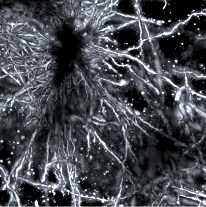
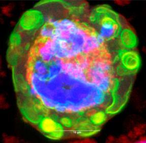
An inverse Colgi-cox stained image of the human brain cortex by Dominical Kaul

Our goal

The Matosin Lab broadly aims to understand how stress contributes to the development of mental illness. The lab has two main streams:

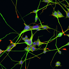
  1. In the first stream, we aim to understand what happens to the cells and molecules in the human brain after stress exposure or in mental illness. To do this, we study human brains donated to science by people who used to live with a mental illness and/or had very stressful lives. Tiny slivers of brain or pieces no larger than the size of a pea are used to pinpoint differences in the shapes, numbers, orientation and connections of brain cells, as well as what is happening inside them from the level of the gene to the protein. This research provides the fundamental knowledge needed to develop new treatments and interventions.
  2. In the second stream, we aim to understand what are the long-term and sustained effects of stress on the human body, and then to build a framework for identifying people who are at risk to mental illness and ways to improve their resilience. Our group is also interested in how the effects of stress and trauma can be transmitted from parent to offspring, therefore having transgenerational impact. To address these questions, we study biological samples  – including saliva, mouth swabs, blood, and breast milk – and psychological data from people and communities who have been heavily stress exposed. By studying human tissues and fluids that are easily accessible and minimally invasive to collect, this research provides the possibility to develop ways to (a) screen for people at risk to the detrimental effects of stress, (b) identify who could benefit from specific treatments and interventions, and (c) design those treatment and interventions.

Our values in and out of the lab

Scientific excellence, impactful research, collaboration, openness and authenticity, training and connecting the next generation of scientists with world leaders, enthusiasm, creating an environment that is positive and encourages teamwork and generosity.

View Dr Natalie Matosin's Scholars page

Contact nmatosin@uow.edu.au for more information.

The Wilson research group is focussed on both basic and applied science relating to chaperones and protein folding, with a special emphasis on a novel group of (normally secreted) extracellular chaperones discovered by us. We reported the first known extracellular chaperone in mammals (clusterin) and have continued to discover new examples of this small but growing family of important molecules. Our studies include in vitro structure-function studies of extracellular chaperones, and also encompass work in small animal models (Drosophila, zebrafish and C. elegans) addressing basic science questions and specific disease scenarios. We have also developed new fluorescence-based technology platforms, including a high-throughput flow cytometry system currently being applied in a search for novel drugs to treat motor neurone disease.

A 3-colour image of a stressed cell: nucleus (blue), endoplasmic reticulum (red) and a chaperone (BiP; green)

View Senior Professor Mark Wilson's Scholars page

Contact mrw@uow.edu.au for more information.

The Yerbury lab is dedicated to understanding the molecular mechanisms underpinning Motor Neurone Disease (MND), with a particular focus on protein misfolding and protein aggregation. Utilising a broad array of methods ranging from the fields of biophysics, biochemistry, and cell and molecular biology, we study the basic biological processes that lead to protein aggregation, with the aim of identifying and developing novel therapeutic strategies for the treatment of MND.

Currently, we are developing a powerful high-throughput microscopy method to screen novel and clinically-approved compounds for their protective properties against MND-related protein aggregation in cultured cells. Utilising this approach, we are able to identify clinically-translatable compounds and assess their therapeutic potential in preclinical models, in an effort to uncover novel treatment avenues for MND.

Motor neurons in culture captured using confocal microscopy (Christen Chisholm, PhD candidate)

View Professor Justin Yerbury's Scholars page

Contact jjyerbury@googlemail.com for more information.