We use cookies to improve your experience on our site and to show you personalised advertising. To find out more, read our privacy policy and cookie policy
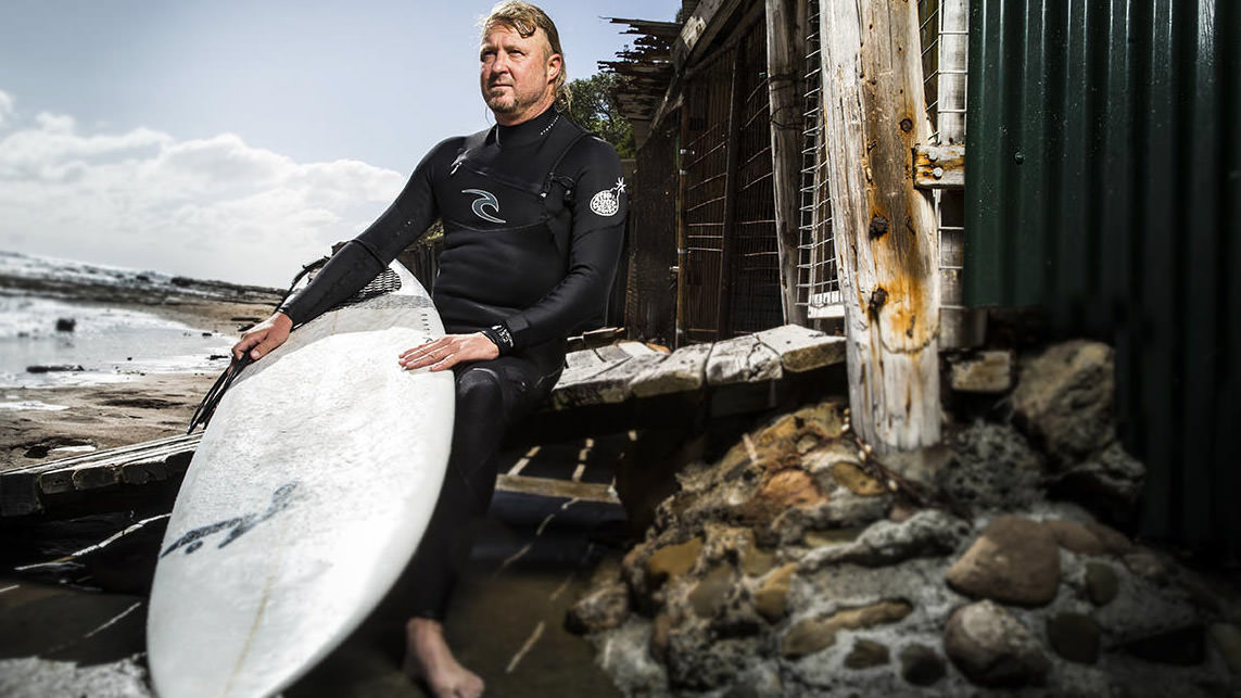
Surfer, scientist, engineer and inventor: James Bouwer has played a key role in developing the microscope technology that is revolutionising our understanding of life at the molecular level.
A new generation of microscopes that allow scientists to look at molecules and proteins down to an atomic resolution has brought about a revolution in molecular biology, opening new doors to fighting disease and to understanding the mechanics of life at the cellular level.
And it's video gamers who we can thank for making it possible.
"Without those guys we wouldn't be here right now," says Dr James Bouwer, General Manager of Cryogenic Electron Microscopy at the University of Wollongong. Driven by the demand for better graphics in video games, the computer chips that render images and video have become massively more powerful and, as a by-product, have helped make cryogenic electron microscopy (cryo-EM) an incredibly powerful tool.
"Things happen in strange ways sometimes," Dr Bouwer says. "So, thank you to all the video game fanatics out there!"
The other key technological development was in direct detection devices - super sensitive cameras - and we can thank Dr Bouwer himself for these: he was a pioneer in this field, designing and building the world's first 64-megapixel camera system, and the world's first direct detection devices, among other things.
"It really is revolutionary ... it’s giving us a real understanding of the mechanics of biology at that molecular resolution." - James Bouwer
In cryo-EM, samples (typically protein molecules) are studied at cryogenic temperatures so that the water in which they are suspended can be frozen in a glass-like state. A beam of high-energy electrons is fired through the sample and then through a lens, generating multiple two-dimensional images that can be converted into three-dimensional images, revealing the molecule's physical structure down to an atomic resolution.
Cryo-EM has been around for decades but the development of these new detectors over the past few years has proved to be a game changer. "The direct detector camera systems are really what's revolutionised cryo-electron microscopy," Dr Bouwer says. "It opened up a whole new world of really high-resolution imaging.
"It has allowed electron microscopes to have enough signal-to-noise ratio to detect every electron that comes down the microscope with a really high signal. Before that you didn't have enough signal and the things you were looking at, the proteins, would just fall apart.
"Thanks to Cryo-EM, molecular science can help us understand the machinery of human cells and how diseases work. We can then develop drugs that influence how these diseases behave to treat or cure them."
[Music]
When I was approached by the University of Wollongong, Judy Raper came to visit me and she suggested maybe I should come out and do a little visit to Australia. My wife and I and more recently, when the children have spent time traveling around the world visiting surf destinations so we love an adventure.
[Music]
The facility that we're building is Molecular Horizons. Molecular Horizons is designed to house three very high-end Transmission Electron Microscopes and an Ion Milling Scanning Electron Microscope. So that project is planned to be completed in 2019. In the interim we've purchased two microscopes so far. The first one has been installed here in Building 41 and this is the Talus Arctica Fei Thermo Fisher development. This microscope is being installed and tested. This microscope we believe will deliver resolution on the order of about 3 angstrom.
So one of the things about Electron Microscopy that's really marvelous is this ability to span the scales that are the living organism. So if you think about a human being, the human is about 2 meters and if you move to the next level down you might be looking at an MRI machine where you might be able to visualize what the brain is doing down to possibly even millimeters. So a thousandth of a meter. If you move from there you might take a light microscope, high and light microscope and start to look at tissues and cells and things inside those cells that would take you down to about a micron which is a thousandth of a millimeter. From there you have to move into an Electron microscope to see beyond that or possibly using x-rays.
So the Electron Microscope enables us to visualize things on the order of millimeters down to microns and beyond a micron. If you divide that up into about a thousand parts you get to a nanometer. Those are sort of the scales of connections in the brain such as axons and things like that. And if you move down another factor of 10 below that you get to the angstrom resolution which is about the diameter of a Hydrogen Atom.
[Music]
Engineering a breakthrough
With his easy-going manner, long blond hair and tan, James Bouwer looks every bit the laid-back Californian surfer that he is. He also happens to be one of the world's leading microscopy experts. When UOW went looking for someone to manage its cryo-EM facility, the key component of the University's new $80 million research centre Molecular Horizons, Dr Bouwer was at the top of its list.
When it's completed, the purpose-built Molecular Horizons Building will house two cryo-electron microscopes: the three-metre tall, one tonne Titan Krios - the world's most powerful high-resolution electron microscope - and the slightly smaller Talos Arctica.
In the meantime, Talos Arctica has been set up in UOW's Sciences Building and begun operations, while Titan Krios will be installed temporarily at the Australian Nuclear Science and Technology Organisation.
The two microscopes will work in tandem, with researchers able to use the smaller, faster Talos Arctica to assess the effectiveness of their experiment before using the Titan Krios.
As well as developing the cameras that revolutionised cryo-EM, Dr Bouwer spent three years managing the University of California, San Diego (UCSD) cryo-EM facility. Before that he had 20 years at the US National Center for Microscopy and Imaging Research (NCMIR) in San Diego designing experiments and running a large suite of microscopes.
"The microscopes … are extremely sensitive; you're trying to locate individual atoms within a larger protein structure. Everything has to be perfect." - James Bouwer
As the NCMIR's principal developmental engineer he also designed automation devices, putting motors on microscopes so they could be operated remotely, and developed software to process the images.
"It was software to create very large montages of tile images, so you could take, say a one millimetre by one millimetre high-resolution image of brain tissue and turn that into a trillion-pixel image - so that one-millimetre square becomes the size of the Google Maps of planet Earth."
When describing the development of his breakthrough four-arm, 64-megapixel camera system, Dr Bouwer sounds like he could be talking about something he knocked together in his backyard shed - until you remember he's talking about incredibly sensitive and extremely expensive equipment.
"We got together with the physicists at the Center for Astrophysics and Space Sciences at UCSD and went and stole some of their particle detectors and bolted them to the bottom of our electron microscopes," he says.
"We started using those as a way to directly bombard the detector so that you got really large signals. From that we developed the direct detector camera systems."
Knowing his love of surfing, when James Bouwer flew to Australia to interview for the job at UOW, the University organised a surf guide to take him to the renowned Sandon Point break.
While other microscopes, using X-ray crystallography, can produce images of a similar resolution, they can't match the speed of cryo-EM and they can't capture the same range of molecules.
"X-ray crystallography is a very high-resolution technique but you have to make protein crystals and that's very difficult," Dr Bouwer says. "People spent 10 years trying to make a crystal of some particular protein and it never happened.
"Now you can just throw those proteins into the electron microscope, take 10,000 pictures of the particles and their structure just falls out for you without having to crystallise. That's the big advantage."
The 2017 Nobel Prize in Chemistry was awarded to Jacques Dubochet, Joachim Frank and Richard Henderson for their role in the development of cryo-electron microscopy, a method which "has moved biochemistry into a new era" the Nobel Committee for Chemistry said. There is a feeling in the scientific community that this Nobel Prize is just the beginning.
Getting it right first time
One of the big attractions for Dr Bouwer in coming to UOW was the opportunity to be involved from the start in setting up a best-practise cryo-EM facility. The numerous surf breaks along the Illawarra coast didn't hurt either. When he flew out to Australia to interview for the role, UOW made sure he got the opportunity to sample those waves.
"The University got me a surf guide who took me to Sandon Point and that was it," he says. "Now my house looks down onto Sandon Point and from my living room I can see the surf. I went and bought some nice big binoculars, so I've got my thumb on the pulse out there."
Dr Bouwer saw in Wollongong the opportunity to use all his experience and knowledge of microscopy, to use the lessons learnt from other facilities around the world, to help UOW get it right first time.
"That was the attraction: to come in at the ground floor and build this into a world-class cryo-electron microscopy centre," Dr Bouwer says.
"I'm working with the designers and architects to make sure we don't have any issues with magnetic fields or vibrations or temperature control. I'm working with the IT people to make sure we have the computing capacity to deal with the data flows coming off the microscopes.
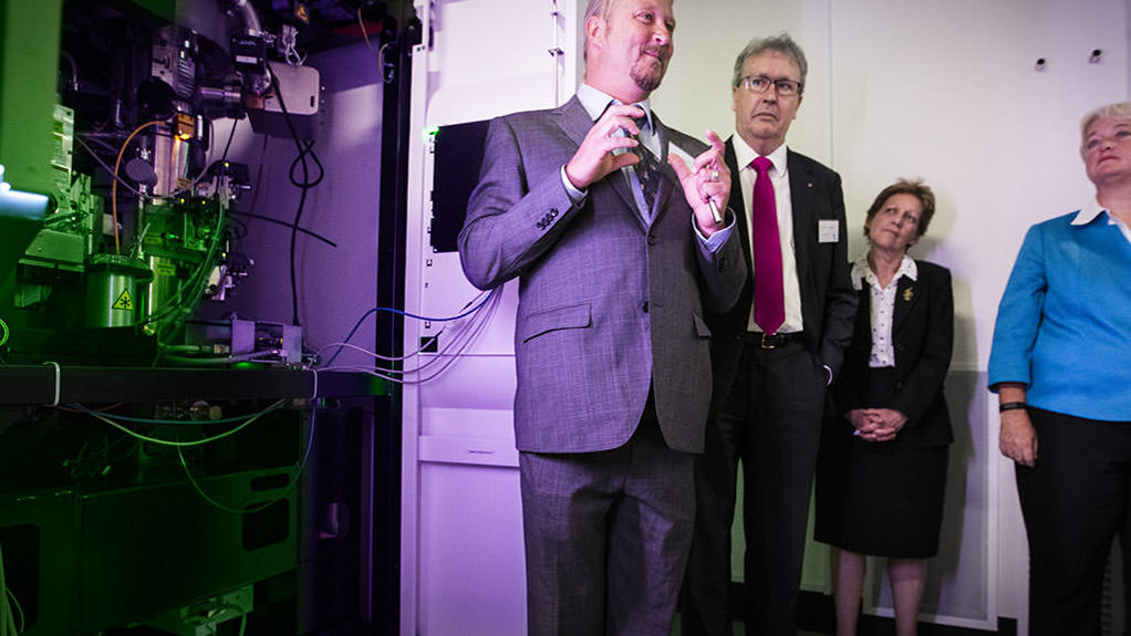
Dr Bouwer explains the inner workings of the Talos Artica cryo-electron microscope to UOW Vice-Chancellor Professor Paul Wellings CBE, Deputy Vice-Chancellor (Research) Professor Judy Raper and Deputy Vic Chancellor (Health and Strategy) Professor Alison Jones.
"The microscopes aren't going to perform properly if you don't take all of that into account. They're extremely sensitive systems; you're looking at atoms essentially, trying to locate individual atoms within a larger protein structure. Everything has to be perfect.
"If there's anything environmentally wrong, it's going to ruin your resolution; things like electromagnetic interference, poor temperature control, the wrong humidity, acoustic issues - just having fan noise can be a problem.
"UOW has made a large effort to meet and exceed all the specifications the microscope manufacturer has laid out. To me that attitude is really nice because what I'm used to when I'm setting up a microscope is they just point and say, 'there's a room over there, put your microscope in there and good luck'.
"It's a different kind of mentality; we really want to get this right. It's a big, ambitious project so I'm really excited to be here helping to make it happen."
Collaboration not competition
The Molecular Horizons building may still be under construction, but the centre is already becoming an Australian hub for molecular life sciences research.
Ever since the Talos Arctica began operations, in December 2017, scientists have been making their way to Wollongong from across the country and around the world. When the Titan Krios is up and running later in 2018 demand for the microscopes will increase further.
This cooperative aspect of the project is something else that Dr Bouwer finds inspiring.
"I'm lucky to have landed in the right spot at the right time and to be here in Wollongong setting this whole thing up and making it happen and turning it into a hub for the whole of Australia," he says.
"Once both microscopes are fully up and running we’ll be putting out four to six terabytes of data every day". - James Bouwer
"One of the attractions of this is all the collaborative efforts and building those relationships. It's a highly collaborative environment because it's so complex, so multidisciplinary, that no one person can know it all."
As well as developing relationships and collaborations with individual researchers and research teams, Molecular Horizons has led to collaborations and partnerships between UOW and other institutions.
"The microscopes require supercomputers on the back end to make things happen," Dr Bouwer says. "Once both microscopes are fully up and running we'll be putting out four to six terabytes of data every day.
"You need a lot of infrastructure - a lot of processing power and a lot of storage - to manage that. Monash University has stepped up to be our partners in the computing side of things, which means we can leverage their massive supercomputer capabilities.
"Cryo-EM and molecular science is not about being a competition. We are striving to build a community of researchers working together to maximise the value of the investments in the technology that UOW and Monash have made."
In this time of unprecedented change, we have a choice to drive change or be driven by it. At the University of Wollongong we choose to lead it and that is why we are investing in an 18 million dollar molecular Life Sciences facility. This world-leading Molecular Science research facility will be dedicated to solving some of the biggest health challenges facing the world today such as developing new forms of Antibiotics, curing Cancer and reversing Alzheimer's disease. The centrepiece of UOW's Molecular Science initiative will be the seven million dollar ultra high-resolution Titan Cryo Krios EM microscope. One of only two in Australia and only a small number across the world. UOW's Molecular Horizons Facility will put New South Wales and in fact Australia on the map as a leader in this revolutionary science and open up unlimited possibilities for vital health-related discoveries.
[Music]
Future focus
While microscope technology will continue to develop and improve, Dr Bouwer says the focus in the field in coming years will be on what cryo-EM can reveal about the mechanics of life.
"The big move now is in things like understanding basic biology at the atomic resolution," he says. "How these little motors turn and ratchet the DNA through. How the little motor proteins walk along microtubules and deliver their cargo loads to various places in the cell.
"And drug development is a really big area; there's a lot of research going into, for example, HIV drugs and how they dock into certain motor proteins to block actions like delivering the DNA to the cell. Or seeing that there's a little pocket in this protein so we can put our drug right in there and block what the protein does.
"The highest resolution I'm getting now is about 1.8 Angstrom resolution on a protein - an Angstrom is about the diameter of a hydrogen atom, so I'm getting resolution down to 1.8 hydrogen atoms.
"Once you get to those levels you can see from the structure what it looks like, what the shape of it is. You can tell what amino acid is sitting there just by the shape that you see in your reconstruction. You don't have to go and try to figure out through old-school chemistry what atom is there, what amino acid it is.
"This is where things are going, and it really is revolutionary. There are hundreds of structures being solved now every year. It's giving us a real understanding of the mechanics of biology at that molecular resolution."